Your Cart is Empty
Shipping is free on orders over €60!
Shipping is free on orders over €60!

Glutathione is the tripeptide γ-L-glutamyl-L-cysteinylglycine. It consists of glutamic acid, cysteine, and glycine. In animal cells, it is the most abundant non-protein thiol, meaning a compound with a free sulfhydryl group. It reaches concentrations in the range of 1 to 10 mmol/l, with the highest levels found in the liver (Meister and Anderson, 1983). It exists in two forms. Reduced glutathione (GSH, reduced glutathione) has a free thiol group on its cysteine residue and is the biologically active form. Oxidised glutathione (GSSG, glutathione disulfide) is formed when two GSH molecules join through a disulfide bridge after donating electrons. In a healthy cell, the reduced form strongly predominates. The ratio of GSH to GSSG therefore serves as a sensitive indicator of oxidative stress (Meister and Anderson, 1983). The term “reduced” on dietary supplements does not mean a lower content. It means that the product contains the active reduced form rather than the disulfide. Glutathione has a wide range of physiological functions. It reacts directly with reactive oxygen species. It serves as a cofactor for glutathione peroxidase (GPx, glutathione peroxidase) in the breakdown of hydrogen peroxide and lipid hydroperoxides. It is a substrate for glutathione-S-transferases (GST, glutathione S-transferases), which conjugate it with substances foreign to the body, including medicines and heavy metals, facilitating their excretion through bile and urine. It participates in the regeneration of vitamins C and E, redox signalling, maintenance of protein thiol groups, and lymphocyte function. It also serves as a cysteine reservoir (Meister and Anderson, 1983). The history of the substance dates back to 1888. The French researcher Joseph de Rey-Pailhade described a sulphur-reducing substance in yeasts and animal tissues and called it “Philothion”. Frederick Gowland Hopkins isolated it in 1921, named it glutathione, and initially considered it a dipeptide (Hopkins, 1921). He did not describe its correct tripeptide composition until 1929 (Hopkins, 1929). Harington and Mead carried out its chemical synthesis in 1935 (Harington and Mead, 1935). We also obtain glutathione from food. Jones and colleagues measured its content in foods listed in the American National Cancer Institute’s questionnaire. Fresh fruit, vegetables, and freshly prepared meat contain substantially more than dairy products, cereals, and canned or otherwise industrially processed foods. Its content also decreases during cooking and storage (Jones et al., 1992). Daily dietary intake is therefore highly variable. Tissue GSH levels decline with age and in numerous chronic diseases. In older people, deficiency is associated with reduced synthesis, which can be corrected by supplying the precursors cysteine and glycine (Sekhar et al., 2011b). A similar pattern has been described in poorly controlled diabetes (Sekhar et al., 2011a). On the dietary supplement market, glutathione is offered as an antioxidant, a “detox” product, support for the liver and immune system, and, in Southeast Asia, for the controversial practice of skin lightening. Injectable, inhaled, and intranasal forms investigated in clinical medicine do not belong to the dietary supplement category. The best-known commercial raw material is “Setria” from the Japanese company Kyowa Hakko Bio, produced through yeast fermentation. In 2018, the company announced self-affirmed generally recognised as safe status (GRAS, generally recognized as safe) for its use in foods at amounts of 5 to 400 mg per serving (Kyowa Hakko USA, 2018). In December 2009, the US Food and Drug Administration (FDA, Food and Drug Administration) completed its review of GRAS Notice No. 293 (GRN 293, GRAS notice) submitted by the Japanese company Kohjin without objections. This concerns the use of glutathione in conventional foods at amounts of 5 to 742.5 mg per serving (FDA, 2009).
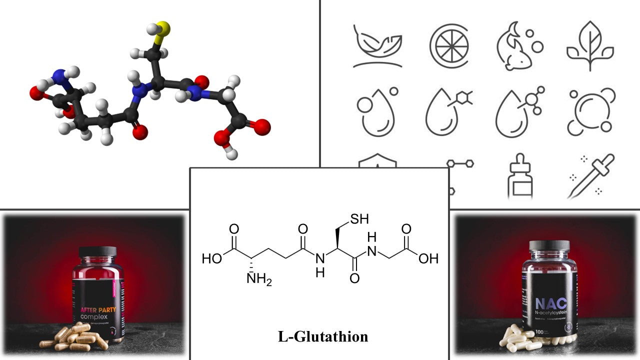
The systematic name of reduced glutathione is (2S)-2-amino-5-[[(2R)-1-(carboxymethylamino)-1-oxo-3-sulfanylpropan-2-yl]amino]-5-oxopentanoic acid. However, the name γ-L-glutamyl-L-cysteinylglycine is generally used. Its molecular formula is C10H17N3O6S, its molar mass is 307.32 g/mol, and its registration number in the Chemical Abstracts Service (CAS) database is 70-18-8 (NCBI, 2026). It is a white crystalline powder. It is readily soluble in water, at approximately 100 g/l, only very slightly soluble in ethanol, and melts with decomposition at temperatures of approximately 185 to 195 °C (Council of Europe, 2022). An unusual feature of the molecule is its γ-glutamyl peptide bond. In this bond, glutamate is linked to cysteine through the carboxyl group of its side chain rather than through the α-carboxyl group, as in proteins (Fig. 1). This bond protects glutathione from ordinary peptidases. It is cleaved almost exclusively by γ-glutamyl transferase (GGT, gamma-glutamyl transferase) on the cell surface, particularly in the kidneys, intestine, and liver (Meister and Anderson, 1983). The reactive centre of the molecule is the thiol group of cysteine. Its pKa is approximately 9. At physiological pH, it is therefore predominantly protonated, and only the smaller thiolate fraction is fully reactive. During a reaction with oxidants, two GSH molecules combine to form GSSG. Glutathione reductase carries out the reverse reduction using nicotinamide adenine dinucleotide phosphate (NADPH, nicotinamide adenine dinucleotide phosphate), supplied by the pentose phosphate pathway. The enzyme contains flavin adenine dinucleotide (FAD, flavin adenine dinucleotide) as a prosthetic group. The functioning of the entire system therefore also depends on an adequate supply of riboflavin. Biosynthesis takes place in two steps, both of which consume adenosine triphosphate (ATP, adenosine triphosphate). First, γ-glutamylcysteine ligase (GCL, glutamate cysteine ligase) joins glutamate to cysteine. This step determines the rate of the entire synthesis and is feedback-inhibited by GSH itself. The limiting factor is usually cysteine availability. Glutathione synthetase (GS, glutathione synthetase) then adds glycine. Breakdown takes place within the γ-glutamyl cycle described by Alton Meister. It begins with the action of GGT and continues with the cleavage of cysteinylglycine by dipeptidases. The released amino acids are then reused by cells for synthesis (Meister and Anderson, 1983). The same GGT enzyme is also why orally administered glutathione is largely broken down in the intestine and liver. Industrially, glutathione is produced mainly through fermentation of the yeasts Saccharomyces cerevisiae or Candida utilis, followed by extraction and crystallisation. Enzymatic synthesis is an alternative. Chemical synthesis from protected amino acids is costly because the thiol group must be protected. In the solid state, the substance is relatively stable. In solution and when exposed to air, however, it gradually oxidises to GSSG. Traces of metal ions, light, higher temperatures, and alkaline pH accelerate oxidation. The raw material is therefore stored in dry, cool conditions at 2 to 8 °C in tightly sealed packaging, with sensitive formulations stored under nitrogen. A prepared solution should be used immediately. The quality of the raw material is defined by the European Pharmacopoeia. Its content is determined by iodometric titration, with a requirement of 98.0 to 101.0 % on a dry-substance basis. Impurities are monitored by capillary electrophoresis, including GSSG, for which a maximum of 1.5 % is permitted (Council of Europe, 2022). In addition to reduced GSH, the market offers oxidised GSSG, S-acetyl-L-glutathione (SAG, S-acetyl glutathione), liposomal and micellar glutathione (Sinha et al., 2018; Solnier et al., 2026), sublingual tablets (Schmitt et al., 2015), and synthesis precursors. These include N-acetylcysteine (NAC, N-acetylcysteine), glycine, and their combination known as “GlyNAC” (Kumar et al., 2023).

Figure 1.L-glutathione (reduced), its structure, natural sources, and related preparations.
The central question regarding glutathione as a dietary supplement is its oral bioavailability. After ingestion, the tripeptide encounters GGT in the intestinal mucosa and liver, which splits it into cysteinylglycine and glutamate. Peptidases then release the individual amino acids. Witschi and colleagues administered a single dose of 0.15 mmol/kg of glutathione, approximately 3 g, to 7 healthy volunteers. During the following 270 minutes, they found no significant increase in plasma glutathione, cysteine, or glutamate. They concluded that the systemic availability of a single dose in humans is negligible (Witschi et al., 1992). The randomised controlled trial (RCT, randomized controlled trial) by Allen and Bradley reached a similar conclusion. In that study, 40 healthy adults took 500 mg twice daily or placebo for 4 weeks. Neither blood glutathione levels nor oxidative stress markers changed (Allen and Bradley, 2011). In rats, however, plasma GSH concentrations doubled from approximately 15 to 30 µmol/l following oral administration. Experiments with inhibitors of synthesis and breakdown showed that this was predominantly due to the absorption of the intact molecule (Hagen et al., 1990). Transport of intact GSH across the intestinal epithelium was also confirmed by in vitro and ex vivo models (Kovacs-Nolan et al., 2014). Park and colleagues administered 50 mg/kg of glutathione to humans. Free GSH in plasma and blood cells did not change, but GSH bound to plasma proteins increased significantly between 60 and 120 minutes (Park et al., 2014). Older studies may therefore have overlooked absorbed glutathione because they measured only the free fraction. The most important human data came from a six-month RCT involving 54 healthy non-smokers. They took 250 or 1000 mg of glutathione daily or placebo. After 6 months, average GSH levels in the higher-dose group increased by 30 to 35 % in erythrocytes, plasma, and lymphocytes and by 260 % in buccal cells. At the lower dose, levels increased by 17 % in whole blood and by 29 % in erythrocytes. The cytotoxicity of natural killer cells (NK, natural killer) in the higher-dose group increased to more than twice that of the placebo group after 3 months (Richie et al., 2015). The study was funded by Kyowa Hakko Bio, which supplied both the tested raw material, “Setria”, and the placebo. It has not yet been independently replicated. A pilot study by the same research group using liposomal glutathione at a dose of 500 or 1000 mg daily for 4 weeks was conducted in 12 adults without a placebo group. It reported an increase in blood GSH of up to 40 % and increased NK-cell cytotoxicity (Sinha et al., 2018). A 2026 crossover pilot study in 14 healthy adults compared single doses of 300 mg of micellar glutathione, 300 mg of liposomal glutathione, and 500 mg of conventional glutathione. The micellar form achieved approximately 2.5 times the exposure of the conventional form (Solnier et al., 2026). However, this was a small study using a patented product, and the result requires independent confirmation. A randomised crossover study in 20 people with metabolic syndrome compared sublingual GSH, conventional oral GSH, and NAC over 3 weeks each. Only the sublingual form improved the plasma ratio of GSH to GSSG (Schmitt et al., 2015). An open-label pilot study included 34 patients with non-alcoholic fatty liver disease (NAFLD, non-alcoholic fatty liver disease), of whom 29 completed the study. After 3 months of lifestyle changes, they received 300 mg of glutathione daily for 4 months, and a reduction in alanine aminotransferase (ALT, alanine aminotransferase) was observed (Honda et al., 2017). However, the absence of a control group prevents the effect of glutathione from being distinguished from that of the lifestyle changes. A double-blind crossover study in 8 healthy men reported less fatigue and lower lactate levels during exercise after two weeks of taking 1 g daily: 2.9 compared with 3.4 mmol/l with placebo. However, some of the authors were employed by the glutathione manufacturer (Aoi et al., 2015). Stronger evidence exists for indirectly supporting synthesis. In 8 older people aged 60 to 75 years, erythrocyte GSH concentrations were approximately half those of 8 young control participants. Two weeks of supplementation with cysteine in the form of NAC and with glycine increased these concentrations by 94.6 % and restored oxidative stress markers to the values found in young people (Sekhar et al., 2011b). A sixteen-week placebo-controlled RCT in 24 older adults using the “GlyNAC” combination, with both glycine and NAC administered at 100 mg/kg daily each, improved glutathione levels, oxidative stress, mitochondrial function, inflammatory markers, and physical performance (Kumar et al., 2023). Vitamin C increased lymphocyte glutathione in people with ascorbate deficiency (Lenton et al., 2003). The second area is skin lightening. The proposed mechanism is inhibition of tyrosinase and a shift in melanin synthesis from dark eumelanin towards lighter pheomelanin (Sonthalia et al., 2016). In an RCT involving 60 medical students, taking 250 mg of glutathione twice daily for 4 weeks reduced the melanin index at all six measured sites. However, the difference compared with placebo was statistically significant at only two sites (Arjinpathana and Asawanonda, 2012). An open-label study without a control group in 30 Filipino women using a lozenge containing 500 mg of glutathione for 8 weeks reported skin lightening (Handog et al., 2016). A three-arm RCT in 60 women compared 250 mg of GSH daily, 250 mg of GSSG daily, and placebo over 12 weeks. A reduction in the melanin index was observed only at some sites, together with a slight improvement in wrinkles (Weschawalit et al., 2017). Reviews highlight small sample sizes, short study durations, and the reversibility of the effect (Sonthalia et al., 2018). A 2025 systematic review summarised five RCT and one open-label study using oral glutathione at doses of 250 mg once or twice daily and 500 mg once daily. All reported a significant reduction in the melanin index compared with placebo. However, the authors consider the effects of both oral and topical glutathione to be only moderately strong and not sustained. They describe intravenous administration as contraindicated because of insufficient efficacy and adverse effects (Sarkar et al., 2025). The Philippine drug authority warned as early as 2011 against intravenous glutathione use outside approved indications, which do not include skin lightening (Sonthalia et al., 2016). In the European Union (EU), this is not a health claim, and a cosmetic effect may not be communicated for a dietary supplement. The third group comprises clinical indications. In Parkinson’s disease, an open-label study in 9 patients reported a 42 % reduction in impairment after intravenous administration of 600 mg twice daily for 30 days (Sechi et al., 1996). However, a randomised, double-blind pilot study in 20 patients using intravenous doses of 1400 mg three times weekly for 4 weeks did not confirm a significant benefit over placebo (Hauser et al., 2009). Intranasal administration of 100 or 200 mg three times daily for 3 months in 45 patients was no better than placebo (Mischley et al., 2017). In cystic fibrosis, inhaled administration of 646 mg twice daily for 6 months in 153 patients did not improve lung function, the number of exacerbations, or quality of life (Griese et al., 2013). Glutathione deficiency has also been described in infection with the human immunodeficiency virus (HIV, human immunodeficiency virus), in diabetes, and during ageing (Sekhar et al., 2011a).
In clinical studies, oral glutathione was administered at doses of 250 to 1000 mg daily for periods ranging from 4 weeks to 6 months (Allen and Bradley, 2011; Richie et al., 2015). The liposomal form was studied at doses of 500 and 1000 mg daily (Sinha et al., 2018), and the micellar form at 600 mg daily for 30 days (Solnier et al., 2026). Studies focusing on the skin used 250 to 500 mg daily (Arjinpathana and Asawanonda, 2012; Weschawalit et al., 2017), while the NAFLD study used 300 mg daily (Honda et al., 2017). In medical applications outside dietary supplements, intravenous doses of 600 to 1400 mg were administered several times weekly (Hauser et al., 2009; Sechi et al., 1996). The effect on the body’s stores develops slowly. At 1000 mg daily, changes in some markers appeared after 1 month. The maximum was reached after 6 months, and levels returned to baseline one month after discontinuation (Richie et al., 2015). Glutathione therefore requires regular, long-term use; an acute effect should not be expected. Commercial preparations usually contain 250 to 500 mg per capsule, with a recommendation of 1 to 2 capsules daily. The highest usual daily dose of 1000 mg thus corresponds to the higher dose used in the six-month RCT. Taking it on an empty stomach is justified by lower digestive peptidase activity, but direct comparative studies are lacking. Combining it with vitamin C has a technological rationale, as ascorbic acid protects the GSH in the formulation against oxidation. The safety profile of oral glutathione is favourable. No serious adverse effects occurred in the six-month RCT (Richie et al., 2015). In the four-week RCT, neither blood counts nor liver and kidney test results changed (Allen and Bradley, 2011). Thirty days of taking 600 mg of the micellar form daily produced no safety signals (Solnier et al., 2026). Indirect support is provided by the European Food Safety Authority’s opinion (EFSA, European Food Safety Authority) on SAG from September 2026. According to this opinion, the safe dose is 300 mg daily for adults and 190 mg and 269 mg daily for younger and older adolescents, respectively, excluding pregnant and breastfeeding women. The panel explicitly stated that human clinical data on orally administered glutathione support its safety in adults (EFSA, 2026). No formal tolerable upper intake level has been established. Reported adverse effects include mild digestive symptoms, such as bloating and looser stools (Allen and Bradley, 2011). Inhaled glutathione caused bronchoconstriction in 8 patients with mild asthma, with a 19 % reduction in forced expiratory volume in one second (FEV1, forced expiratory volume in one second). The authors attributed this to the formation of sulphites (Marrades et al., 1997). People with asthma and those sensitive to sulphites should therefore exercise caution, particularly with inhaled forms. The claim that long-term glutathione use lowers zinc levels could not be substantiated in the peer-reviewed literature. Among interactions, cancer treatment is the most frequently discussed. In an RCT involving 50 patients with gastric cancer, intravenous glutathione reduced cisplatin neurotoxicity without diminishing the antitumour response. After 15 weeks, neuropathy occurred in 4 of 24 patients, compared with 16 of 18 in the placebo group (Cascinu et al., 1995). In 151 women with ovarian cancer treated with cisplatin, it reduced toxicity and improved quality of life (Smyth et al., 1997). Pharmacokinetic interactions with common medicines have not been described. Given its limited absorption, significant drug interactions are not expected. Data are lacking for pregnant and breastfeeding women and for children, so use is not recommended.
In practice, reduced glutathione should be viewed as one of several ways to support the body’s own glutathione system. The best-supported precursor is NAC. It supplies cysteine as the limiting substrate for synthesis and is the standard antidote for paracetamol poisoning. In the Czech Republic, it is authorised as a medicine, for example as a mucolytic under the trade name ACC. In some EU countries, it is sold in dietary supplements, but its regulatory status differs from country to country. The combination of glycine and NAC known as “GlyNAC” is supported by a placebo-controlled RCT in older adults (Kumar et al., 2023). This is complemented by the smaller studies in older people and people with diabetes described above (Sekhar et al., 2011a; Sekhar et al., 2011b). However, the samples are small, and the results originate mainly from a single research group. Whey protein is naturally rich in cysteine and may support synthesis through nutrition. Alpha-lipoic acid participates in the regeneration of reduced forms of antioxidants, including GSH. Vitamin C is the only one of these substances with direct human evidence, as it increases lymphocyte glutathione in people with vitamin C deficiency (Lenton et al., 2003). Selenium is an essential component of glutathione peroxidase, and riboflavin, as a precursor of FAD, supplies the cofactor required by glutathione reductase. Sulforaphane from broccoli activates the transcription factor Nrf2 (nuclear factor erythroid 2 related factor 2), which regulates GCL expression, among other processes. Silymarin from milk thistle (Silybum marianum) is traditionally used to support the liver. However, the systematic Cochrane review of 13 randomised studies involving 915 patients did not confirm a significant effect on mortality or complications in alcohol-related and viral liver diseases (Rambaldi et al., 2007). Among the modified forms, S-acetylglutathione is the most important, and its safety was assessed favourably by EFSA (EFSA, 2026). However, placing it on the EU market requires authorisation as a novel food. So far, liposomal and micellar glutathione have only pilot data without placebo controls (Sinha et al., 2018; Solnier et al., 2026), while the sublingual form has only one small crossover study (Schmitt et al., 2015).
Written for Kratom World by:
Ing. Matěj Malík, Ph.D.
Research Scientist, Postdoctoral Researcher
Faculty of Agrobiology, Food and Natural Resources, Czech University of Life Sciences Prague
Department of Agroenvironmental Chemistry and Plant Nutrition

References
Allen, J., Bradley, R.D., 2011. Effects of oral glutathione supplementation on systemic oxidative stress biomarkers in human volunteers. Journal of Alternative and Complementary Medicine. 17, 827-833. https://doi.org/10.1089/acm.2010.0716.
Aoi, W., Ogaya, Y., Takami, M., Konishi, T., Sauchi, Y., Park, E.Y., Wada, S., Sato, K., Higashi, A., 2015. Glutathione supplementation suppresses muscle fatigue induced by prolonged exercise via improved aerobic metabolism. Journal of the International Society of Sports Nutrition. 12, 7. https://doi.org/10.1186/s12970-015-0067-x.
Arjinpathana, N., Asawanonda, P., 2012. Glutathione as an oral whitening agent: a randomized, double-blind, placebo-controlled study. Journal of Dermatological Treatment. 23, 97-102. https://doi.org/10.3109/09546631003801619.
Cascinu, S., Cordella, L., Del Ferro, E., Fronzoni, M., Catalano, G., 1995. Neuroprotective effect of reduced glutathione on cisplatin-based chemotherapy in advanced gastric cancer: a randomized double-blind placebo-controlled trial. Journal of Clinical Oncology. 13, 26-32. https://doi.org/10.1200/JCO.1995.13.1.26.
EFSA, Panel on Nutrition, Novel Foods and Food Allergens (NDA), 2026. Safety of S-acetyl glutathione as a novel food pursuant to Regulation (EU) 2015/2283. EFSA Journal. 24, e10258. https://doi.org/10.2903/j.efsa.2026.10258.
FDA, 2009. GRAS Notice No. GRN 000293, Glutathione. Notifier Kohjin Co., Ltd. U.S. Food and Drug Administration, Silver Spring. https://hfpappexternal.fda.gov/scripts/fdcc/index.cfm?id=293&set=grasnotices.
Griese, M., Kappler, M., Eismann, C., Ballmann, M., Junge, S., Rietschel, E., van Koningsbruggen-Rietschel, S., Staab, D., Rolinck-Werninghaus, C., Mellies, U., Köhnlein, T., Wagner, T.O., König, S., Teschler, H., Heuer, H.-E., Kopp, M., Heyder, S., Hammermann, J., Küster, P., Honer, M., Mansmann, U., Beck-Speier, I., Hartl, D., Fuchs, C., Hector, A., 2013. Inhalation treatment with glutathione in patients with cystic fibrosis. A randomized clinical trial. American Journal of Respiratory and Critical Care Medicine. 188, 83-89. https://doi.org/10.1164/rccm.201303-0427OC.
Hagen, T.M., Wierzbicka, G.T., Sillau, A.H., Bowman, B.B., Jones, D.P., 1990. Bioavailability of dietary glutathione: effect on plasma concentration. American Journal of Physiology. 259, G524-G529. https://doi.org/10.1152/ajpgi.1990.259.4.G524.
Handog, E.B., Datuin, M.S.L., Singzon, I.A., 2016. An open-label, single-arm trial of the safety and efficacy of a novel preparation of glutathione as a skin-lightening agent in Filipino women. International Journal of Dermatology. 55, 153-157. https://doi.org/10.1111/ijd.12999.
Harington, C.R., Mead, T.H., 1935. Synthesis of glutathione. Biochemical Journal. 29, 1602-1611. https://doi.org/10.1042/bj0291602.
Hauser, R.A., Lyons, K.E., McClain, T., Carter, S., Perlmutter, D., 2009. Randomized, double-blind, pilot evaluation of intravenous glutathione in Parkinson's disease. Movement Disorders. 24, 979-983. https://doi.org/10.1002/mds.22401.
Honda, Y., Kessoku, T., Sumida, Y., Kobayashi, T., Kato, T., Ogawa, Y., Tomeno, W., Imajo, K., Fujita, K., Yoneda, M., Kataoka, K., Taguri, M., Yamanaka, T., Seko, Y., Tanaka, S., Saito, S., Ono, M., Oeda, S., Eguchi, Y., Aoi, W., Sato, K., Itoh, Y., Nakajima, A., 2017. Efficacy of glutathione for the treatment of nonalcoholic fatty liver disease: an open-label, single-arm, multicenter, pilot study. BMC Gastroenterology. 17, 96. https://doi.org/10.1186/s12876-017-0652-3.
Hopkins, F.G., 1921. On an autoxidisable constituent of the cell. Biochemical Journal. 15, 286-305. https://doi.org/10.1042/bj0150286.
Hopkins, F.G., 1929. On glutathione: a reinvestigation. Journal of Biological Chemistry. 84, 269-320. https://doi.org/10.1016/S0021-9258(18)77062-2.
Jones, D.P., Coates, R.J., Flagg, E.W., Eley, J.W., Block, G., Greenberg, R.S., Gunter, E.W., Jackson, B., 1992. Glutathione in foods listed in the National Cancer Institute's Health Habits and History Food Frequency Questionnaire. Nutrition and Cancer. 17, 57-75. https://doi.org/10.1080/01635589209514173.
Kovacs-Nolan, J., Rupa, P., Matsui, T., Tanaka, M., Konishi, T., Sauchi, Y., Sato, K., Ono, S., Mine, Y., 2014. In vitro and ex vivo uptake of glutathione (GSH) across the intestinal epithelium and fate of oral GSH after in vivo supplementation. Journal of Agricultural and Food Chemistry. 62, 9499-9506. https://doi.org/10.1021/jf503257w.
Kumar, P., Liu, C., Suliburk, J., Hsu, J.W., Muthupillai, R., Jahoor, F., Minard, C.G., Taffet, G.E., Sekhar, R.V., 2023. Supplementing Glycine and N-Acetylcysteine (GlyNAC) in Older Adults Improves Glutathione Deficiency, Oxidative Stress, Mitochondrial Dysfunction, Inflammation, Physical Function, and Aging Hallmarks: A Randomized Clinical Trial. The Journals of Gerontology: Series A. 78, 75-89. https://doi.org/10.1093/gerona/glac135.
Kyowa Hakko USA, 2018. Kyowa Hakko USA announces GRAS self-affirmation for Setria glutathione. Press release dated 7 February 2018. https://www.prweb.com/releases/kyowa_hakko_usa_announces_gras_self_affirmation_for_setria_glutathione/prweb15182463.htm.
Lenton, K.J., Sané, A.T., Therriault, H., Cantin, A.M., Payette, H., Wagner, J.R., 2003. Vitamin C augments lymphocyte glutathione in subjects with ascorbate deficiency. American Journal of Clinical Nutrition. 77, 189-195. https://doi.org/10.1093/ajcn/77.1.189.
Marrades, R.M., Roca, J., Barberà, J.A., de Jover, L., MacNee, W., Rodriguez-Roisin, R., 1997. Nebulized glutathione induces bronchoconstriction in patients with mild asthma. American Journal of Respiratory and Critical Care Medicine. 156, 425-430. https://doi.org/10.1164/ajrccm.156.2.9611001.
Meister, A., Anderson, M.E., 1983. Glutathione. Annual Review of Biochemistry. 52, 711-760. https://doi.org/10.1146/annurev.bi.52.070183.003431.
Mischley, L.K., Lau, R.C., Shankland, E.G., Wilbur, T.K., Padowski, J.M., 2017. Phase IIb study of intranasal glutathione in Parkinson's disease. Journal of Parkinson's Disease. 7, 289-299. https://doi.org/10.3233/JPD-161040.
NCBI, 2026. PubChem Compound Summary for CID 124886, Glutathione. National Center for Biotechnology Information, Bethesda. https://pubchem.ncbi.nlm.nih.gov/compound/124886.
Park, E.Y., Shimura, N., Konishi, T., Sauchi, Y., Wada, S., Aoi, W., Nakamura, Y., Sato, K., 2014. Increase in the protein-bound form of glutathione in human blood after the oral administration of glutathione. Journal of Agricultural and Food Chemistry. 62, 6183-6189. https://doi.org/10.1021/jf501338z.
Council of Europe, 2022. Glutathione. European Pharmacopoeia, 11th edition. European Directorate for the Quality of Medicines & HealthCare, Strasbourg.
Rambaldi, A., Jacobs, B.P., Gluud, C., 2007. Milk thistle for alcoholic and/or hepatitis B or C virus liver diseases. Cochrane Database of Systematic Reviews. 4, CD003620. https://doi.org/10.1002/14651858.CD003620.pub3.
Richie, J.P., Nichenametla, S., Neidig, W., Calcagnotto, A., Haley, J.S., Schell, T.D., Muscat, J.E., 2015. Randomized controlled trial of oral glutathione supplementation on body stores of glutathione. European Journal of Nutrition. 54, 251-263. https://doi.org/10.1007/s00394-014-0706-z.
Sarkar, R., Yadav, V., Yadav, T., P, J., Mandal, I., 2025. Glutathione as a skin-lightening agent and in melasma: a systematic review. International Journal of Dermatology. 64, 992-1004. https://doi.org/10.1111/ijd.17535.
Schmitt, B., Vicenzi, M., Garrel, C., Denis, F.M., 2015. Effects of N-acetylcysteine, oral glutathione (GSH) and a novel sublingual form of GSH on oxidative stress markers: A comparative crossover study. Redox Biology. 6, 198-205. https://doi.org/10.1016/j.redox.2015.07.012.
Sechi, G., Deledda, M.G., Bua, G., Satta, W.M., Deiana, G.A., Pes, G.M., Rosati, G., 1996. Reduced intravenous glutathione in the treatment of early Parkinson's disease. Progress in Neuro-Psychopharmacology and Biological Psychiatry. 20, 1159-1170. https://doi.org/10.1016/S0278-5846(96)00103-0.
Sekhar, R.V., McKay, S.V., Patel, S.G., Guthikonda, A.P., Reddy, V.T., Balasubramanyam, A., Jahoor, F., 2011a. Glutathione synthesis is diminished in patients with uncontrolled diabetes and restored by dietary supplementation with cysteine and glycine. Diabetes Care. 34, 162-167. https://doi.org/10.2337/dc10-1006.
Sekhar, R.V., Patel, S.G., Guthikonda, A.P., Reid, M., Balasubramanyam, A., Taffet, G.E., Jahoor, F., 2011b. Deficient synthesis of glutathione underlies oxidative stress in aging and can be corrected by dietary cysteine and glycine supplementation. American Journal of Clinical Nutrition. 94, 847-853. https://doi.org/10.3945/ajcn.110.003483.
Sinha, R., Sinha, I., Calcagnotto, A., Trushin, N., Haley, J.S., Schell, T.D., Richie, J.P., 2018. Oral supplementation with liposomal glutathione elevates body stores of glutathione and markers of immune function. European Journal of Clinical Nutrition. 72, 105-111. https://doi.org/10.1038/ejcn.2017.132.
Smyth, J.F., Bowman, A., Perren, T., Wilkinson, P., Prescott, R.J., Quinn, K.J., Tedeschi, M., 1997. Glutathione reduces the toxicity and improves quality of life of women diagnosed with ovarian cancer treated with cisplatin: results of a double-blind, randomised trial. Annals of Oncology. 8, 569-573. https://doi.org/10.1023/A:1008211226339.
Solnier, J., Du, M., Zhang, Y., Roh, Y.S., Kuo, Y.C., Ibi, A., Wood, S., Hardy, M., Gahler, R.J., Chang, C., 2026. A targeted metabolomic assessment of oral glutathione bioavailability and safety in humans: a randomized crossover clinical trial. Antioxidants. 15, 354. https://doi.org/10.3390/antiox15030354.
Sonthalia, S., Daulatabad, D., Sarkar, R., 2016. Glutathione as a skin whitening agent: Facts, myths, evidence and controversies. Indian Journal of Dermatology, Venereology and Leprology. 82, 262-272. https://doi.org/10.4103/0378-6323.179088.
Sonthalia, S., Jha, A.K., Lallas, A., Jain, G., Jakhar, D., 2018. Glutathione for skin lightening: a regnant myth or evidence-based verity? Dermatology Practical & Conceptual. 8, 15-21. https://doi.org/10.5826/dpc.0801a04.
Weschawalit, S., Thongthip, S., Phutrakool, P., Asawanonda, P., 2017. Glutathione and its antiaging and antimelanogenic effects. Clinical, Cosmetic and Investigational Dermatology. 10, 147-153. https://doi.org/10.2147/CCID.S128339.
Witschi, A., Reddy, S., Stofer, B., Lauterburg, B.H., 1992. The systemic availability of oral glutathione. European Journal of Clinical Pharmacology. 43, 667-669. https://doi.org/10.1007/BF02284971.